Cellular Analysis Core
Histology Core Sign Up Calendar: Google Calendar Histology
Flow Core Sign Up Calendar: Google Calendar Flow Core.
Fluorofinder Account (Detailed laser/detector configurations): https://fluorofinder.com/ . Select "Montana State University".
The Cellular Analysis Facility (CAF) in the Department of Microbiology and Cell Biology, located in the Health Sciences Building, is focused on the analysis of the phenotype and function of cells and tissues by flow cytometry, histology, and single cell transcriptomics (scRNA Seq). We provide instrumentation including FACS analyzers, a FACS sorter, an imaging cytometer, a tissue processor and embedder, microtomes and cryostats, fluorescent microscopes, and a 10x Genomics Chromium Controller, as well as user training and core services. Facility Director Diane Bimczok, DVM, PhD, can be reached at 406-994-4928 or diane.bimczok@montana.edu
The Flow Core Facility is located in the Health Sciences Building, HSB 247 and HSB 241. There are also three instruments in Cooley #29.
The histology core facility is located in the Health Sciences Building #263A.
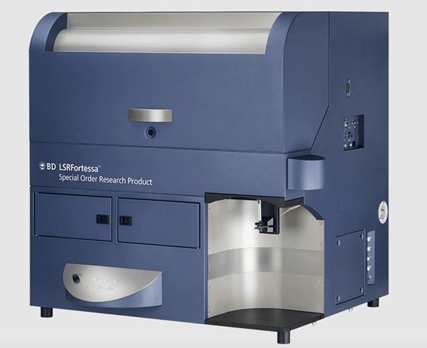
Three of the flow cytometers are located in the Cooley building (room 29) for easy access by researchers on campus: BD Calibur, Stratedigm, and BD Fortessa.
For more information contact Andy Sebrell at 406-994-2878 or andysebrell@montana.edu.
Core Facility Instrumentation
Please acknowledge the following award when publishing any data generated with the BioRad ZE5 Cell Analyzer: USDA 2023-70410-41180.
ZE5™ Cell Analyzer
The new ZE5 is equipped with five lasers and filters to analyze up to 27 colors, plus forward and side scatter. The ZE5™ has a fully integrated, automated sample loader, compatible with all standard tubes and culture plates; has a specialized small particle (20 nm to 1 µm) analyzer for microbiology, virology and exosome research.

Features of the ZE5 Cell Analyzer: Sample loading system:
- Universal sample loader: easily switch between tubes, 96-well plates and 384-well plates with no hardware changes using the integrated sample loader.
- Temperature control and orbital agitation
- Sample pump with minimal dead volume and sample recovery feature
- Automated plate reader can read a 96-well plate in 15 minutes and a 384-well plate in 60 minutes
- Can analyze up to 100,000 cells per second
Stratedigm SE520EON
Please cite the following funding source in any meeting, abstract, poster, talk and publication if the Stratedigm Cytometer was used to generate data: Montana NIH INBRE P20GM103474.
The SE520EON is a 2-laser (488 nm with 530, 580, 676 and 780 nm detectors and 640 nm with 676 and 780 nm detectors), 6-color fixed configuration system.
BD LSR Fortessa

Purchased with funding from the M.J. Murdock Charitable Trust in 2014, the Fortessa represents a high dimensional analyzer, capable of analyzing 18 colors plus forward and side scatter, for a total of 20 parameters. It is equipped with a 640 nm laser (with detectors for 670, 780 and 790 nm), a 561 nm laser (with detectors for 582, 660, 780, 610 and 710 nm), a 488 nm laser (with detectors for 695 and 590 nm, FSC and SSC), a 405 nm laser (with detectors for 670, 525, 710, 610 and 450 nm) and a 355 nm UV laser (with detectors for 450 and 525 nm). Each excitation source is supported by polygon detector arrays, and each polygon can support up to eight detectors for maximum flexibility in optical configuration. The BD Fortessa is located in the Cooley building and provides high-dimensional FACS capacity to research groups on campus. The Fortessa was recently repaired with funding from MCB and is now back in full working order.
BD FACS Caliburs
Two FACS Calibur analyzers are available for basic cell analysis, with one instrument located in the HSB FACS Core room and a second one in the Cooley building (Cooley 29). The FACS Caliburs are equipped with two lasers (488 nm and 642 nm), allowing the analysis of up to 6 different cell parameters
BD FACS Aria II cell sorter
The FACS Aria is a high-end 8- to10-parameter flow cytometer with high-speed cell sorting capability. The FACS Aria is contained within a biosafety cabinet allowing for sorting of BSL-2 level-infected or primary human samples.

Detailed laser/detector configurations for each cytometer may be found at https://fluorofinder.com/. Select "Montana State University."
ImageStream imaging cytometer
Imaging cytometry (ImageStream) combines features of flow cytometry and high-resolution light and fluorescent microscopy analysis of cell suspensions to capture images for each event passing through the detector in a flow stream. The ImageStream ISX Mach II imaging cytometeris equipped with three lasers (488/642/785 nm) to record six image channels per object (bright field, dark field and 5 fluorescence channels), a multiple magnification option (20x, 40x and 60x) to image both eukaryotic cells and bacteria.

10X Genomics Single cell RNA sequencing
Single Cell Sequencing (scRNASeq) is as a novel, state-of-the-art approach that enables comprehensive analyses of heterogeneous cell populations and that is revolutionizing transcriptomics studies.
The 10x Genomics Chromium Controller is a user-friendly droplet-based scRNA-seq system that enables processing of up to 80,000 individual cells in up to 8 samples for downstream RNA sequencing in one single experiment. The Chromium Controller uses drop-based microfluidics to label individual cells in a suspension with genetic barcodes. This barcoding process enables researchers to identify all cDNA sequences from a single cell upon sequencing, based on their unique molecular identifiers. The Chromium platform also is compatible with other single-cell, sequence-based analysis approaches such as studies of chromatin accessibility, cell surface proteins, immune clonotype, antigen specificity, and CRISPR edits.

10X Genomics provides a free, easy-to-use analysis and visualization software specifically designed for the complex datasets generated with the Chromium scRNASeq workflow that enables researchers to access, explore and interpret their data.

|
Flow Cytometry* |
FACS Aria II Sorter – sorting service |
$50/h |
|
|
FACS Aria II Sorter – user operated |
$30/h |
|
|
ImageStream |
$35/h |
|
|
ZE5 Cell Analyzer |
$35/h |
|
|
Stratedigm SE520EON |
$30/h |
|
|
BD CytoFlex# (Under Repair) |
$30/h |
|
|
BD FACS Calibur |
$20/h |
|
Histology |
Processing |
$4/cassette |
|
|
Embedding station |
$1/cassette |
|
|
Microtome |
$15/h |
|
|
Cryostat |
$20/h |
|
|
Staining station (deparaffinizing and H & E) |
$10/h |
|
scRNASeq |
Chromium Controller |
$30/run |
Histology Facility
The histology core facility provides histological services using standard histological techniques. The histology core is part of Microbiology and Cell Biology and is located in the Health Sciences Building #263A. The core can provide paraffin and frozen sections, Immuohistochemistry, processing, embedding, and routine staining of tissues by the staff (fees provided below). With approved training, individual lab personnel may also use the equipment for a fee.

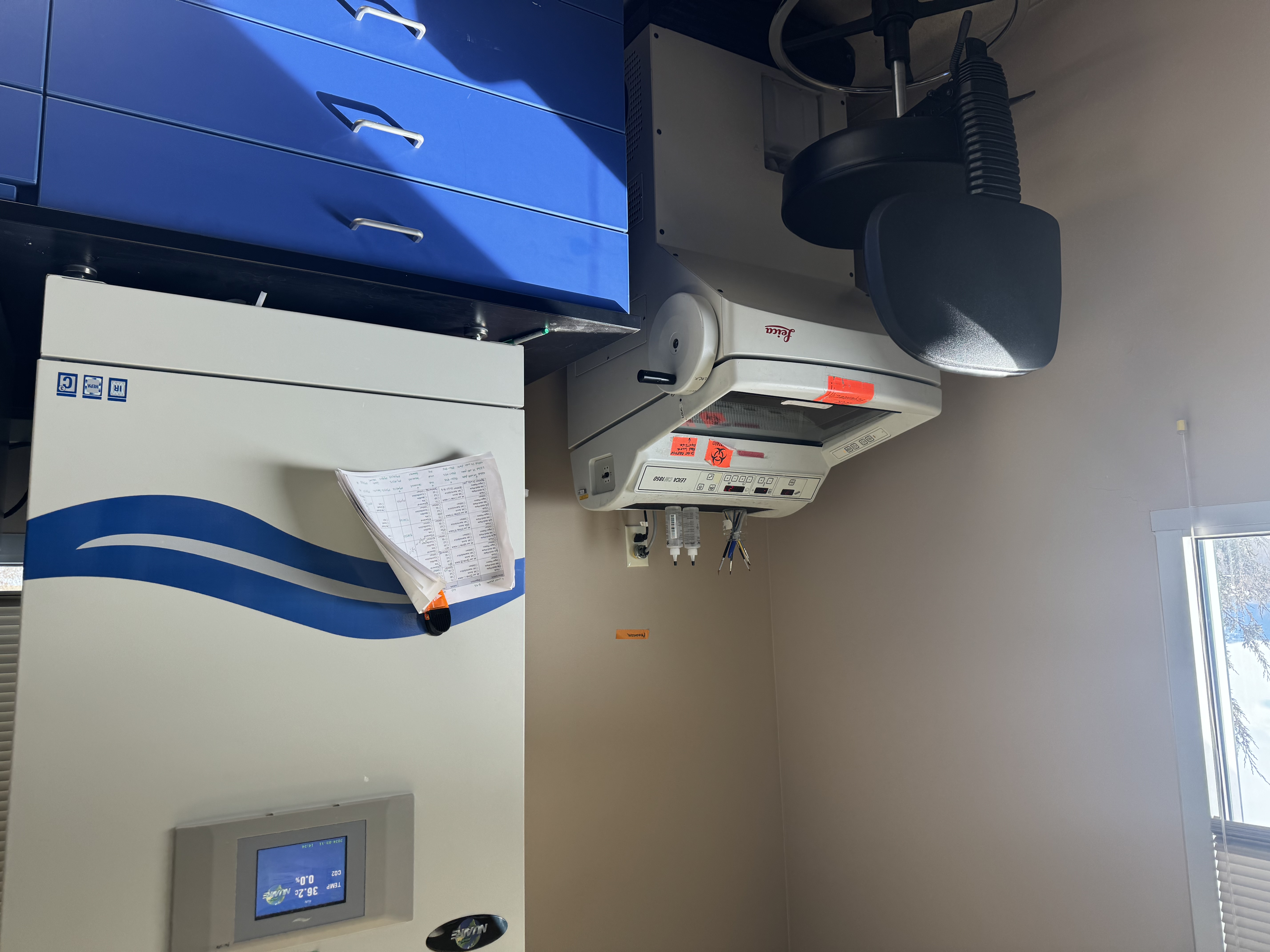
The histology lab has a Leica cryostat for sectioning frozen sections and a VIP6 processor which dehydrates tissues for paraffin sections.
Histopathology Fees
Paraffin Processing:
Tissues submitted to the lab in 70% ETOH (no Formalin, NBF) in labeled cassette or container if you don’t have cassettes. Histology lab will make sure the appropriate size cassette is used before processing. If processing very small samples such as mouse eyes, Organoids, or similar sized tissues please make sure to use cassettes with smaller vents. Tissues will shrink and can be pulled through the vents if too large while processing.
Tissue submitted in 70% ETOH, processed and embedded $6.00 per cassette
Processing $4.00 per cassette
Embedding $2.00 per cassette
Unstained Section on one slide $1.50 per slide
H & E stained slide $2.50 per slide
TRAP stained slide (PI supplies the reagents) $4.00 per slide
Toluidine Blue Stained slide $2.50 per slide
Giemsa, Masons Trichrome $10.00 per slide
PAS TUNEL Stain (PI supplies reagents) $6.00 per slide
Special requests for stains if dyes must be ordered -cost dependent Price TBD
Immunohistochemistry stains requiring enzyme digestion or $12.50 per slide
Antigen Retrieval of paraffin embedded tissue. (PI supplies Antibodies)
Usage of Facility equipment and stations:
Processing $4.00 per cassette
Embedding station $1.00 per cassette
Microtome $15.00 per hour
Cryostat $20.00 per hour
Staining station (deparaffinizing and H & E) $10.00 per hour
Frozen Sectioning:
Snap freezing tissues $5.00 per block
Unstained frozen section $1.50 per slide
H&E stained frozen section $2.50 per slide
Bone Histotechniques:
Decalcification 15% Formic Acid $1.50 per tissue/cassette
Decalcification EDTA $2.50 per tissue/cassette

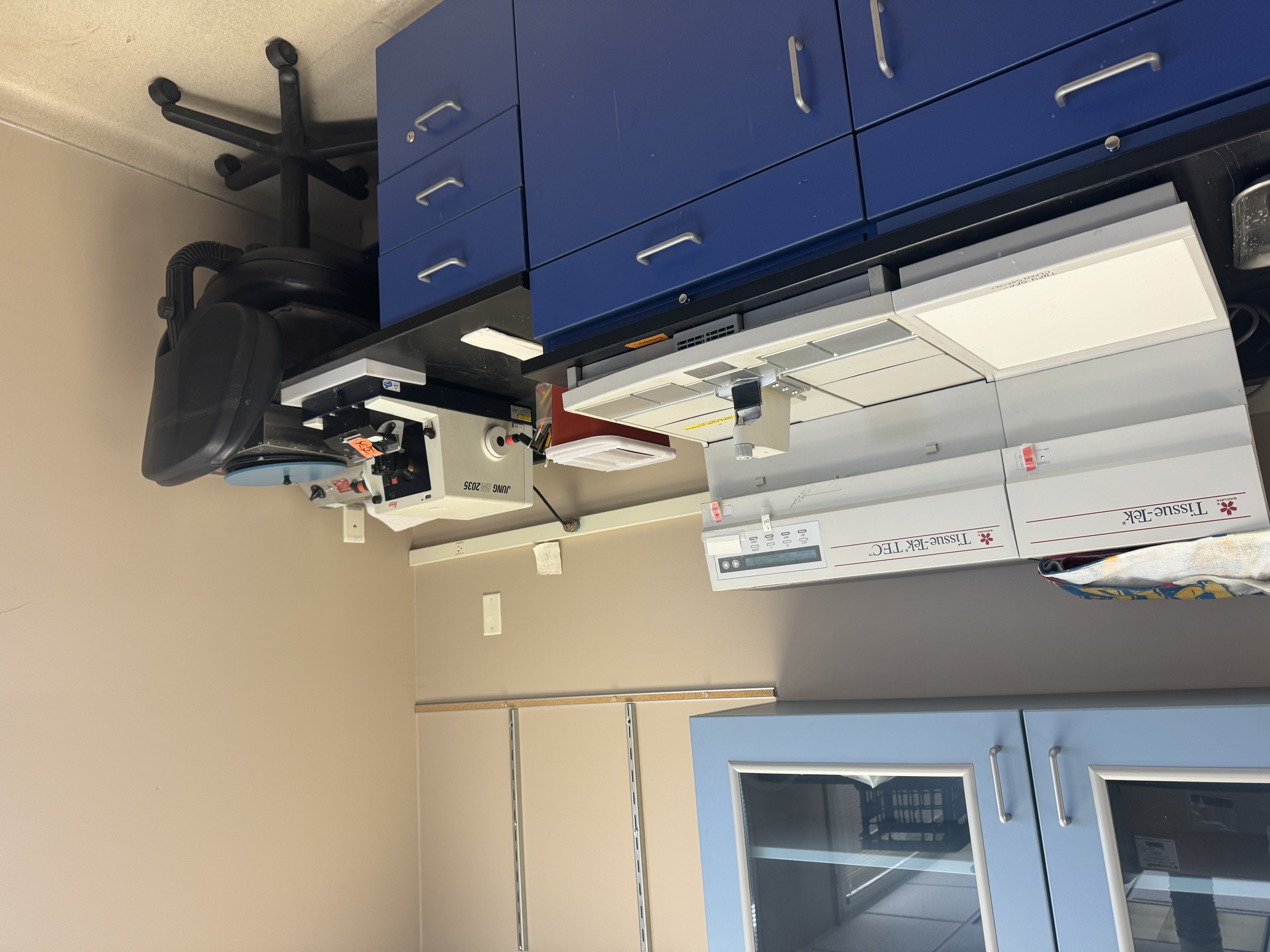
The lab has a Leica microtome for sectioning paraffin embedded tissue. There is also a Tissue Tek paraffin embedding station.

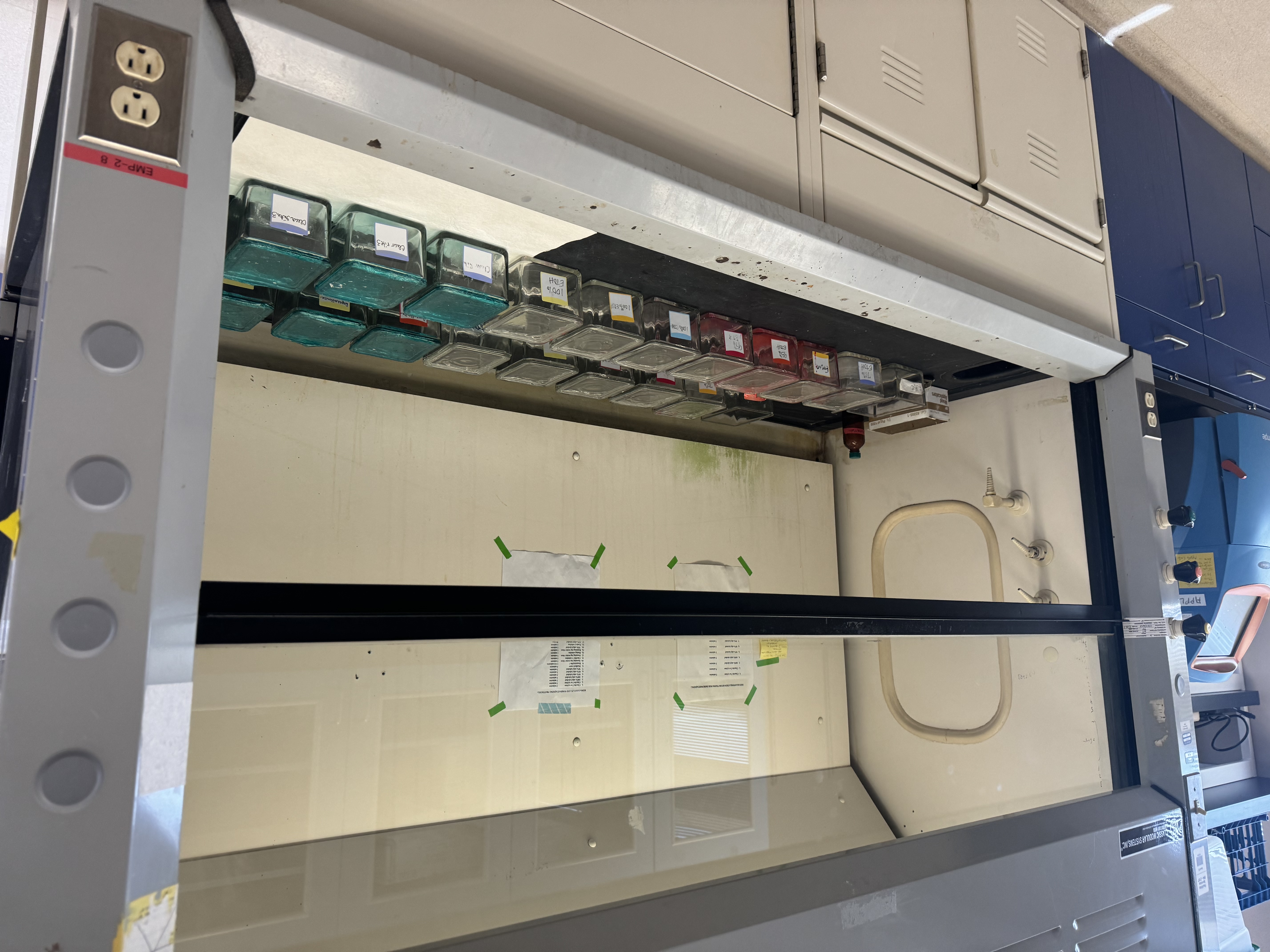
Histology lab has a slide staining station set up for immediate use.
For more information contact Andy Sebrell at 406-994-2878 or andysebrell@montana.edu or Diane Bimczok at 406-994-4928, diane.bimczok@montana.edu.
